Diabetes can affect your sight.
Diabetic Retinopathy is the leading cause of preventable blindness in the United States in people of working age 20 to 65. Severe vision loss can usually be prevented with early diagnosis and treatment. Patients with both insulin dependent and non-insulin dependent diabetes mellitus should have yearly dilated eye exams.
Diabetic retinopathy is a progressive disease, and careful monitoring can usually prevent severe vision loss. Patients with diabetes mellitus do not process and store sugar properly so that high sugar levels cause retinal blood vessel damage (diabetic retinopathy).
Request an AppointmentTypes of diabetic retinopathy.
There are two types of diabetic retinopathy: non-proliferative diabetic retinopathy (NPDR) and proliferative diabetic retinopathy (PDR).
- NPDR is an early stage of diabetic retinopathy. In this stage, small blood vessels called capillaries form microaneurysms within the retina that leak blood or fluid. The leaking fluid causes retinal swelling, deposition of fatty exudates, and blurred vision. Many people with diabetes have mild NPDR, which may not yet affect their vision. It is at this stage that early treatment can be the most effective. When vision is affected, it is the result of macular edema and/or macular ischemia.
- Macular edema is swelling or thickening of the macula, a small area in the center of the retina that is needed for fine visual acuity (the center or bull’s eye of vision). The swelling is caused by fluid leaking from damaged retinal blood vessels. It is the most common cause of visual loss in diabetes. Vision loss may be mild to severe, but even in the worst cases, peripheral vision continues to function.
- Macular ischemia occurs when small blood vessels close. Diabetes causes this problem in many organs increasing the risk for stroke and heart attack. In the retina, vision blurs because the macula no longer receives sufficient blood supply to work properly.
- PDR is present when abnormal new vessels (neovascularization) begin growing on the surface of the retina or optic nerve. The main cause of PDR is inadequate blood flow to the retinal periphery due to closure of retinal blood vessels. The retina responds by growing new blood vessels but, unfortunately rather than replacing the old closed vessels, new blood vessels develop on the surface of the retina and are abnormal, frail, and cause unwanted scar tissue. The scar tissue may cause wrinkling or traction detachment of the retina. PDR may cause more severe vision loss than NPDR because it can affect both central and peripheral vision.
- Vitreous hemorrhage: The fragile new vessels may bleed into the vitreous, a clear, gel-like substance that fills the center of the eye. If the vitreous hemorrhage is small, a person might see only a few new dark floaters. A very large hemorrhage might block out all vision. It may take days or months for the blood to resolve. If the eye does not clear the vitreous blood adequately within a reasonable time, vitrectomy surgery may be recommended. Vitreous hemorrhage alone does not cause permanent vision loss. When the blood clears, vision may return to its former level unless the macula is damaged.
- Traction retinal detachment: When PDR is present, scar tissue associated with neovascularization can shrink, wrinkle, and pull the retina from its normal position. This may occur spontaneously or following laser treatment. Macular wrinkling can cause visual distortion. More severe vision loss can occur if the macula or large areas of the retina are detached.
- Neovascular glaucoma: Occasionally, extensive retinal vessel closure will cause new, abnormal blood vessels to grow on the iris (colored part of the eye) and block the normal flow of fluid out of the eye. Pressure in the eye increases, resulting in neovascularglaucoma, a severe, usually painful, eye disease that causes damage to the optic nerve and can lead to total blindness.
How is diabetic retinopathy diagnosed?
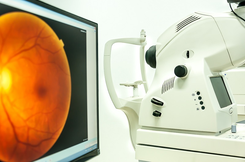
A dilated eye examination is the only way to detect changes inside your eye. An eye doctor can often diagnose and treat serious retinopathy before you are aware of any vision problems. If diabetic retinopathy is diagnosed, specialized testing – including:
- Standard retinal fundus photography (FP)
- Optical Coherence Tomography (OCT)
- Fluorescein Angiography (FA)
– are all utilized to help diagnose, guide treatment, and determine visual prognosis.
How is diabetic retinopathy treated?
Prevention of the development of retinopathy is most important. Strict control of your blood sugar will significantly reduce the long-term risk of vision loss from diabetic retinopathy. Multiple studies have confirmed that the tighter the control of blood sugar the less likely retinopathy will develop or if already present to progress to requiring treatment. If high blood pressure and kidney problems are present, they need to be treated.
Laser Surgery
Laser surgery is often recommended for people with macular edema, PDR, and neovascular glaucoma. For macular edema, a focal or grid laser is focused on the damaged retina near the macula to decrease the fluid leakage. The main goal of treatment is to prevent further loss of vision and to stabilize vision.
For PDR, the laser is focused on all parts of the retina except the macula. This panretinal photocoagulation (PRP) treatment causes abnormal new vessels to shrink and often prevents them from growing in the future. It also decreases the chance that vitreous bleeding or retinal detachment will occur. Multiple laser treatments over time are sometimes necessary. Laser surgery does not cure diabetic retinopathy and does not always prevent further loss of vision.
Intraocular injections
Both patients with NPDR/macular edema and PDR can sometimes benefit from injections of steroids and/or anti-blood vessel medications. These injections can help potentiate grid laser and PRP and aid in vitrectomy surgery when given preoperatively.
Vitrectomy
In advanced PDR, the doctor may recommend a vitrectomy. If vitreous bleeding (hemorrhage) is present the doctor may wait for a month or more to see if the blood clears on its own before performing a vitrectomy or may elect to operate more urgently if rapid visual restoration is necessary (such as with one-eyed patients who are temporarilty blinded by the blood). During this microsurgical procedure, which is performed in the operating room, the blood-filled vitreous is removed and replaced with a clear solution. Vitrectomy often prevents further bleeding by removing the abnormal vessels that caused the bleeding. If the retina is detached, it can be repaired during the vitrectomy surgery. Surgery can sometimes be technically challenging if the patient has extensive tractional changes.
Vision loss is largely preventable.
If you have diabetes, it is important to know that today, with improved methods of diagnosis and treatment, only a small percentage of people who develop retinopathy have serious vision problems. Early detection of diabetic retinopathy is the best protection against loss of vision. You can significantly lower your risk of vision loss by maintaining strict control of your blood sugar and visiting your doctor regularly. The doctor may ask about your current hemoglobin A1C, which is a reliable marker reflecting recent diabetic control success.
When to Schedule an Examination
People with diabetes should schedule examinations at least once a year. More frequent medical eye examinations may be necessary after a diagnosis of diabetic retinopathy. Pregnant diabetic women should schedule an appointment in the first trimester because retinopathy can progress quickly during pregnancy. Rapid changes in blood sugar can cause fluctuating vision in both eyes even if retinopathy is not present.
You should have your eyes checked promptly if you have visual changes that
- affect only one eye,
- last more than a few days, or
- are not associated with a change in blood sugar.
When you are first diagnosed with diabetes, you should have your eyes checked
- within five years of the diagnosis if you are 29 years old or younger, or
- within a few months of the diagnosis if you are 30 years old and older.
Southern Vitreoretinal Associates, P.L. have been managing patients with diabetic retinopathy for 25 years and the doctors are using the latest techniques to help maintain good vision and prevent blindness. Your retinal surgeon or their staff can answer any questions you may have concerning Diabetic Retinopathy.